
Transthoracic Echocardiogram
(also called ECHO)
Introduction
An echocardiogram uses ultrasound waves (signals) to take pictures of your heart. It is used to check your beating heart and its various parts including valves (doors), walls, pumping of blood in and outside heart, Pressures in various parts of heart.
This test provides very important information to your cardiologist to identify any problems in heart. This is a non-invasive and painless test with quick results
There are various types of Echocardiograms (details are given below)
Procedure
An ECHO is a non-invasive test. This test is simple and can be performed at your doctor's office, a clinic or a hospital room. It involves ECHO machine with a screen (like TV and transducer which will obtain images from your hear.
You will be asked to lie down in a bed after expose the chest. The technician will attach sticky patches (electrodes) to your body to help detect and conduct the electrical currents of your heart. The technician will dim the lights of the room to view the images of the heart. Small gel will be applied to your chest in order to get good quality images. The technician will move then move the transducer back and forth over your chest. The images will be recorded in various angles on a monitor, which are recorded for your doctor to review. You may hear a pulsing "whoosh," which the ultrasound is recording the blood flowing through your heart.
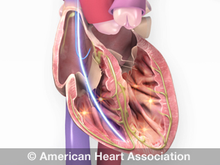
What we look for in HEART during ECHO?
- Heart size
- Pumping strength
- Damage to the heart muscle
- Valve problems
- Heart defects.
Who is ECHO for?
- The ECHO is recommended in the following conditions
- Heart attack or Suspected heart attack
- Palpitations
- Syncope- blackouts
- Heart failure
- Stroke
- Heart valve disease
- Pulmonary Hypertension ( lungs related heart disease)
- Others -** Can be requested by physicians in any medical conditions
How long does the procedure last?
This procedure may take upto 30-40 minutes depends on the images. Sometimes they may use some contrast (coloured dye) to obtain images, therefore the procedure may need extra time.
Types of Echocardiogram
- Transthoracic Echocardiogram- Routine ( as above)
- Trans-esophageal echocardiogram
- Stress echocardiogram
- Doppler echocardiogram